- 1. Mufarrıj IK, Keettel WC. Prolapse of the uterus associated with pregnancy. Am J Obstet Gynecol. 1957; 73: 899–903.
- 2. Hill PS. Uterine prolapse complicating pregnancy. A case report. J Reprod Med. 1984; 29: 631–3.
- 3. Matsumoto T, Nishi M, Yokota M, Ito M. Laparoscopic treatment of uterine prolapse during pregnancy. Obstet Gynecol. 1999; 93: 849.
- 4. Te Linde’s Operative Gynecology, 8th edition. Nonsurgical correction of defects, the use of vaginal support devices. Patricia J. Sulak. Chapter F, pp. 1082–3.
- 5. Guariglia L, Carducci B, Botta A, Ferrazzani S, Caruso A. Uterine prolapse in pregnancy. Gynecol Obstet Invest. 2005; 60: 192–4.
- 6. Klawans AH, Kanter AE. Prolapse of the uterus and pregnancy. Am J Obstet Gynecol. 1949; 57: 939–46.
- 7. Meydanli MM, Ustun Y, Yalcın OT. Pelvic organ prolapse complicating third trimester pregnancy. A case report. Gynecol Obstet Invest. 2005; 61: 133–4.
- 8. Meydanli MM, Ustun Y, Yalcil OT. Pelvic organ prolapse complicating third trimester pregnancy. Gynecol Obstet Invest. 2006; 61: 133–134.
Successful Management of Uterine Prolapse During Pregnancy with Vaginal Pessary at University of Gondar Comprehensive Specialized Referral Hospital, Gondar Ethiopia

Affiliations
1University of Gondar Comprehensive Specialized Hospital, Ethopia.
*Corresponding Author: Dr. Seidomer Abdu Ahmed*
Citation: Seidomer Abdu Ahmed. Successful Management of Uterine Prolapse During Pregnancy with Vaginal Pessary at University of Gondar Comprehensive Specialized Referral Hospital, Gondar Ethiopia. Collect J Gynecol Obstet. Vol 2 (2) 2025; ART0083.
Abstract
Background: Uterine prolapse during pregnancy is a rare condition that may lead to complications such as infection, miscarriage, or preterm labor. Conservative management with a vaginal pessary can help support the uterus and avoid surgical intervention. This case report highlights the successful use of a pessary for managing uterine prolapse during the second trimester.
Methods: A 29-year-old gravida II para I woman presented at 17 weeks and 1 day of gestation with a one-week history of a protruding vaginal mass. Physical examination confirmed second-degree uterine prolapse. An 85 mm ring pessary was inserted. The patient was followed every 2–4 weeks for pessary care and monitoring of potential complications.
Results: Throughout the pregnancy, the pessary remained well-positioned with no signs of infection or discomfort. At 39 weeks, the patient had a spontaneous vaginal delivery of a healthy neonate. Postpartum evaluation at six weeks showed complete resolution of the prolapse with no recurrence or pelvic floor symptoms.
Conclusion
Vaginal pessary use is a safe, effective, and non-invasive method for managing uterine prolapse during pregnancy. With proper follow-up and care, it can help ensure favorable maternal and neonatal outcomes. Conservative treatment should be considered as a first-line option, especially in resource-limited settings.
Keywords: Uterine Prolapse, Pregnancy, Vaginal Pessary.
Introduction
Uterine prolapse during pregnancy is a rare clinical condition, with an estimated incidence of 1 in 10,000 to 15,000 deliveries [1]. It may occur de novo or worsen during pregnancy, particularly in women with preexisting pelvic floor weakness. In most cases, pregnancy exerts an adverse effect on a preexisting prolapse. However, significant uterine prolapse persisting into the third trimester is extremely rare [2]. As the uterus enlarges with advancing gestation, it typically becomes supported by the pelvic brim, which may reduce the degree of prolapse. Despite this, complications such as maternal discomfort, cervical desiccation and ulceration, urinary tract infections, acute urinary retention, spontaneous abortion, preterm labor, fetal or maternal sepsis, and even maternal death can occur [3]. Management options range from conservative approaches, such as bed rest and pessary insertion, to surgical interventions including laparoscopic suspension procedures [4]. The use of vaginal pessaries in pregnancy was first reported in 1949 and remains a valuable non-invasive treatment, especially in resource-limited settings. Among the available options, the silicone-coated ring pessary is particularly favorable due to ease of insertion and self-care by the patient [5]. This report describes a case of uterine prolapse complicating a second-trimester pregnancy that was successfully managed with a vaginal pessary, resulting in a full-term vaginal delivery.
Case Report
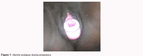
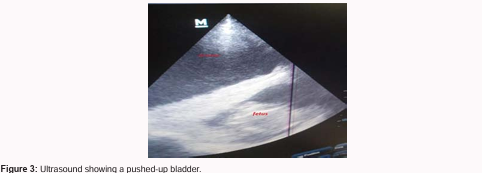
A 29-year-old gravida II para I woman was referred to our hospital at 17 weeks and 1 day of gestation with a one-week history of a protruding vaginal mass. She also reported urinary retention of four days’ duration, for which a urinary catheter had been inserted prior to referral. There was no prior history of uterine prolapse, pelvic trauma, chronic cough, stress urinary incontinence, or other risk factors. Her previous delivery was a spontaneous vaginal birth four years earlier, which was uneventful. On pelvic examination, a 5 × 4 cm globular mass was observed protruding from the vaginal introitus. The cervix, which was closed and not ulcerated, was identified as the leading part of the prolapse and was entirely outside the vagina (Figure 1). Obstetric ultrasound confirmed a viable intrauterine pregnancy consistent with 17+1 weeks of gestation, with normal amniotic fluid volume. The urinary bladder was noted to be distended and displaced above the uterus (Figure 3).
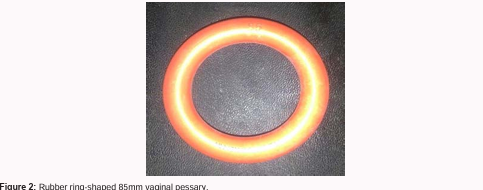
The prolapsed mass was manually reduced, and a vaginal pack was inserted for 24 hours. Subsequently, a ring-shaped silicone vaginal pessary (85 mm) was placed (Figure 2). The patient was educated on pessary self-care and observed in the ward for 48 hours to monitor for urinary or bowel complications. Following pessary insertion, the cervix remained fully intravaginal, and the patient reported no urinary or bowel issues. The patient was followed every two weeks up to 22 weeks of gestation, during which the pessary was removed, cleaned, and reinserted at each visit. From 22 to 36 weeks, she was followed monthly, then weekly until delivery. The remainder of the pregnancy was uneventful, with no complaints of pessary displacement, vaginal irritation, or ulceration. Notably, after 22 weeks of gestation, the prolapse did not recur even when the pessary was temporarily removed during follow-up visits.At 37 weeks and 4 days, the patient had a spontaneous vaginal delivery of a healthy male neonate weighing 3000 grams, with Apgar scores of 7 and 9 at one and five minutes, respectively. She was discharged on the second postpartum day, and the puerperium was uneventful. On follow-up at two months postpartum, there was no clinical evidence of uterine prolapse.
Discussion
Uterine prolapse during pregnancy is a rare clinical condition. Its possible etiologies include prior childbirth trauma, a history of difficult deliveries or macrosomia, congenital connective tissue disorders, obesity, and... Increased intra-abdominal pressure. Physiologic changes of pregnancy, such as cervical elongation, hypertrophy, and ligamentous relaxation due to hormonal effects (particularly progesterone and relaxin), may also contribute to prolapse [5]. In our patient, there was no history of pelvic trauma or obstetric complications, making physiologic changes of pregnancy the most likely cause. While congenital connective tissue disorders could not be ruled out, further evaluation was not possible due to limited diagnostic resources. Management strategies for uterine prolapse in pregnancy range from conservative approaches to surgical interventions. Conservative management includes gynecologic hygiene, bed rest, and positioning in slight Trendelenburg, and has been reported to be effective in some cases [5]. The use of vaginal pessaries, first described in 1949 by Klawans and Kanter, remains a common non-surgical option, particularly in cases with later-onset prolapse [6]. Vaginal pessaries are readily available and easy to apply, though they may be associated with complications such as increased vaginal discharge, odor, mucosal erosion, and urinary retention [4]. In our case, a properly fitted 85 mm ring pessary was used successfully without any complications. Contrary to some reports in the literature that describe frequent pessary dislodgment, our patient retained the pessary throughout the follow-up period with no issues. This success may be attributed to appropriate pessary selection, correct sizing, and patient adherence—key factors in successful conservative management. When conservative options fail or prolonged bed rest is not feasible, laparoscopic uterine suspension may be considered during early pregnancy. However, this technique requires skilled hands, and failed cases have been reported [3]. In women who have completed childbearing, a definitive surgical option such as cesarean hysterectomy with abdominal sacrocolpopexy may be appropriate [7]. Ultimately, management should aim to minimize complications such as discomfort, urinary tract infections, urinary retention, cervical laceration, preterm labor, and maternal or fetal infection and death. In this case, we successfully managed uterine prolapse conservatively using a vaginal pessary, avoiding these complications.
Conclusion
The management of uterine prolapse during pregnancy should be individualized based on patient preference, clinical severity, and available resources. Vaginal pessary use is a safe and effective option that can help prevent maternal and fetal complications and should be included in counseling and management planning.